 5′ to 3′ explained
YouTube channel
Show notes
Why Does Life Need Both DNA and RNA? (DNA vs RNA Animation)
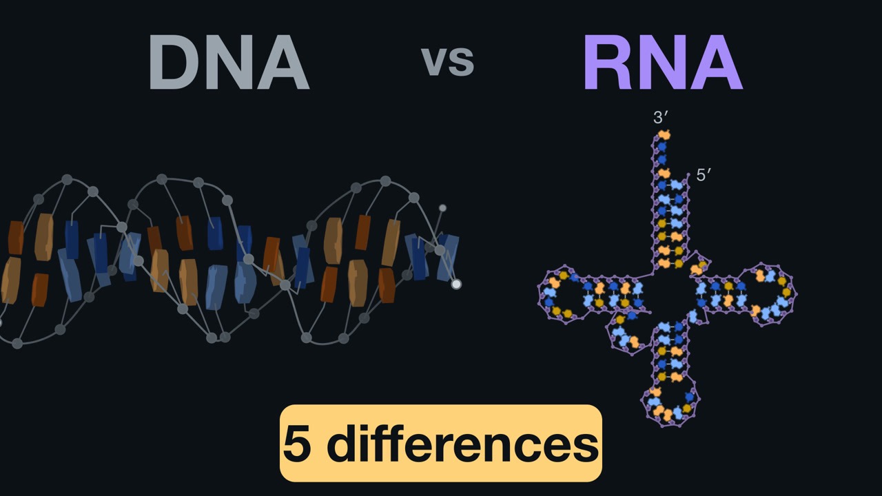
Every living cell uses both DNA and RNA. This video goes through the five differences between them: three in the chemistry (the sugar, the strands, the letters) and two in what that chemistry is for (how long each lasts, and the job each does), and why each difference fits its job.
The video is coming soon to the channel.
Vocabulary
In the order the video explains them.
- DNA (deoxyribonucleic acid) 0:57
- The cell's permanent record of its genes: kept for the cell's whole life and handed on when it divides. Its sugar is deoxyribose.
- RNA (ribonucleic acid) 0:57
- The cell's working copy of a gene's message, written when needed and broken down after use; some RNAs fold into shapes that do chemical work. Its sugar is ribose.
- Nucleotide 1:09
- The building block of DNA and RNA: a sugar, a phosphate and a base (the letter). Nucleotides join into a chain.
- Base 1:09
- The part of a nucleotide that carries the information: the letter (A, G, C, and T in DNA or U in RNA).
- Ribose 1:16
- RNA's sugar. It carries an OH group (an oxygen and a hydrogen) on its 2′ carbon.
- 2′ carbon ("two prime") 1:16
- The second carbon of the sugar ring. The prime mark (′) numbers the sugar's carbons, to tell them apart from the atoms of the base.
- OH group (hydroxyl) 1:16
- An oxygen bonded to a hydrogen. On RNA's 2′ carbon it sits beside the link to the next nucleotide and can attack it, cutting the chain.
- Deoxyribose 1:25
- DNA's sugar: ribose missing the oxygen on its 2′ carbon ("deoxy" = without oxygen). The D in DNA.
- Phosphate 1:34
- The part of a nucleotide that links one sugar to the next, forming the chain's backbone.
- Alkali 1:44
- A strongly basic solution, such as lye. RNA falls apart in it; DNA's links hold.
- Base pairing 2:04
- Letters on two strands pairing across: A with T (or U in RNA), G with C.
- Double helix 2:04
- The shape of DNA's two paired strands, wound around each other.
- Single strand 3:21
- One chain with no partner strand. RNA is usually single stranded: it is written from one of a gene's two strands and is not copied to keep.
- Thymine (T) 3:32
- DNA's fourth letter. It is uracil with one extra methyl group, and it pairs with A.
- Uracil (U) 3:32
- RNA's fourth letter, in place of thymine. It pairs with A. In DNA, a U usually means a damaged cytosine.
- Methyl group 3:42
- One carbon with three hydrogens (CH₃). The extra piece that makes thymine out of uracil.
- Cytosine (C) 4:41
- One of the four letters in both DNA and RNA; it pairs with G.
- Amino group 4:41
- A nitrogen with hydrogens (NH₂). When water knocks it off a cytosine and leaves an oxygen, the cytosine becomes uracil.
- Mismatch 5:00
- Two letters across from each other that are not partners, such as a U across from a G.
- Mutation 5:23
- A change in the DNA's sequence that is kept and copied: here, a C-G pair that has become a T-A pair.
- Repair enzyme (uracil-DNA glycosylase) 5:33
- An enzyme that slides along DNA hunting for uracil, flips it out into a pocket and cuts the base off its sugar, leaving the backbone whole; other enzymes then put back a C.
- Nucleus 7:09
- The compartment of a plant or animal cell where most of its DNA is kept.
- Ribosome 8:25
- The machine that builds every protein. It is made of RNA and protein, but the spot where it joins amino acids together is RNA alone.
- Amino acid
- The building block of proteins; the ribosome joins amino acids into a chain.
- RNA world
- The idea, held by many biologists, that early life ran on RNA, which can both carry information and do chemistry, and that DNA evolved later as a safer place to keep the genes.
Study frame
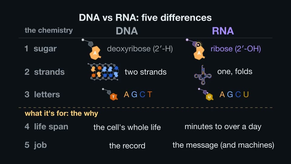
The whole picture in one labelled frame, to save or print.
Review questions
Which carbon of the sugar carries the extra oxygen in RNA, and why does that oxygen make RNA less stable than DNA?
Answer
The 2′ carbon. Its OH group sits next to the link to the next nucleotide and can attack the phosphate, cutting RNA's own chain. DNA's sugar lacks that oxygen, so its chain is far tougher.
Why can a damaged letter in DNA be repaired accurately?
Answer
DNA has two paired strands, so every letter has a partner across from it. The partner strand shows which letter belongs at the damaged spot.
What does cytosine become when it loses its amino group, and what happens if the damage is not repaired before the DNA is copied?
Answer
It becomes uracil, which pairs with A instead of G. After two rounds of copying, the original C-G pair has become a T-A pair: a mutation.
What is the leading idea for why DNA uses T instead of U?
Answer
If DNA used U as a normal letter, a real U and a damaged C would look the same, and the repair enzyme could not tell them apart. With T as the normal letter (marked by its methyl group), any U in DNA is a mistake and safe to cut out.
Name the five differences between DNA and RNA.
Answer
The sugar (deoxyribose vs ribose), the strands (two vs one), the letters (T vs U), the life span (the cell's whole life vs minutes to more than a day), and the job (the record vs the message, and sometimes the machine).
What fact about the ribosome supports the RNA world idea?
Answer
The spot where the ribosome joins amino acids is made of RNA alone: RNA, not protein, does that chemistry. RNA can carry information and do chemistry.
Chapters
- 0:00 Five differences, and why
- 0:33 Two jobs
- 1:09 The sugar
- 2:05 The strands
- 3:33 The letters
- 7:09 Life span
- 7:39 Job: RNA at work
- 9:13 An RNA world?
- 9:35 Five differences
Sources
39 sources. Each was checked against its record (Crossref, PubMed, the Protein Data Bank, or the textbook edition) and against the claim it supports.
- Li Y, Breaker RR (1999). Kinetics of RNA degradation by specific base catalysis of transesterification involving the 2′-hydroxyl group. Journal of the American Chemical Society 121:5364–5372. doi.org/10.1021/ja990592p
- Schroeder GK, Lad C, Wyman P, Williams NH, Wolfenden R (2006). The time required for water attack at the phosphorus atom of simple phosphodiesters and of DNA. Proceedings of the National Academy of Sciences USA 103:4052–4055. doi.org/10.1073/pnas.0510879103
- van der Valk T, Pečnerová P, Díez-del-Molino D, et al. (2021). Million-year-old DNA sheds light on the genomic history of mammoths. Nature 591:265–269. doi.org/10.1038/s41586-021-03224-9
- Watson JD, Crick FHC (1953). Molecular structure of nucleic acids: a structure for deoxyribose nucleic acid. Nature 171:737–738. doi.org/10.1038/171737a0
- Pauling L, Corey RB (1956). Specific hydrogen-bond formation between pyrimidines and purines in deoxyribonucleic acids. Archives of Biochemistry and Biophysics 65:164–181. doi.org/10.1016/0003-9861(56)90185-0
- Carreras CW, Santi DV (1995). The catalytic mechanism and structure of thymidylate synthase. Annual Review of Biochemistry 64:721–762. doi.org/10.1146/annurev.bi.64.070195.003445
- Krokan HE, Drabløs F, Slupphaug G (2002). Uracil in DNA – occurrence, consequences and repair. Oncogene 21:8935–8948. doi.org/10.1038/sj.onc.1205996
- Parikh SS, Mol CD, Slupphaug G, Bharati S, Krokan HE, Tainer JA (1998). Base excision repair initiation revealed by crystal structures and binding kinetics of human uracil-DNA glycosylase with DNA. The EMBO Journal 17:5214–5226. doi.org/10.1093/emboj/17.17.5214
- Mol CD, Izumi T, Mitra S, Tainer JA (2000). DNA-bound structures and mutants reveal abasic DNA binding by APE1 and DNA repair coordination. Nature 403:451–456. doi.org/10.1038/35000249
- Sawaya MR, Prasad R, Wilson SH, Kraut J, Pelletier H (1997). Crystal structures of human DNA polymerase β complexed with gapped and nicked DNA: evidence for an induced fit mechanism. Biochemistry 36:11205–11215. doi.org/10.1021/bi9703812
- Tumbale PP, Jurkiw TJ, Schellenberg MJ, Riccio AA, O'Brien PJ, Williams RS (2019). Two-tiered enforcement of high-fidelity DNA ligation. Nature Communications 10:5431. doi.org/10.1038/s41467-019-13478-7
- Mol CD, Arvai AS, Slupphaug G, et al. (1995). Crystal structure and mutational analysis of human uracil-DNA glycosylase: structural basis for specificity and catalysis. Cell 80:869–878. doi.org/10.1016/0092-8674(95)90290-2
- Lindahl T (1974). An N-glycosidase from *Escherichia coli* that releases free uracil from DNA containing deaminated cytosine residues. Proceedings of the National Academy of Sciences USA 71:3649–3653. doi.org/10.1073/pnas.71.9.3649
- Poole A, Penny D, Sjöberg BM (2001). Confounded cytosine! Tinkering and the evolution of DNA. Nature Reviews Molecular Cell Biology 2:147–151. doi.org/10.1038/35052091
- Schoenberg DR (2011). Mechanisms of endonuclease-mediated mRNA decay. WIREs RNA 2:582–600. doi.org/10.1002/wrna.78
- Narsai R, Howell KA, Millar AH, O'Toole N, Small I, Whelan J (2007). Genome-wide analysis of mRNA decay rates and their determinants in *Arabidopsis thaliana*. The Plant Cell 19:3418–3436. doi.org/10.1105/tpc.107.055046
- Jinek M, Chylinski K, Fonfara I, Hauer M, Doudna JA, Charpentier E (2012). A programmable dual-RNA-guided DNA endonuclease in adaptive bacterial immunity. Science 337:816–821. doi.org/10.1126/science.1225829
- Sternberg SH, Redding S, Jinek M, Greene EC, Doudna JA (2014). DNA interrogation by the CRISPR RNA-guided endonuclease Cas9. Nature 507:62–67. doi.org/10.1038/nature13011
- Nishimasu H, Ran FA, Hsu PD, et al. (2014). Crystal structure of Cas9 in complex with guide RNA and target DNA. Cell 156:935–949. doi.org/10.1016/j.cell.2014.02.001
- Shi H, Moore PB (2000). The crystal structure of yeast phenylalanine tRNA at 1.93 Å resolution: a classic structure revisited. RNA 6:1091–1105. doi.org/10.1017/S1355838200000364
- Nissen P, Hansen J, Ban N, Moore PB, Steitz TA (2000). The structural basis of ribosome activity in peptide bond synthesis. Science 289:920–930. doi.org/10.1126/science.289.5481.920
- Ko D, Ruonala R, Faille A, et al. (2026). Recruitment of bifunctional regulator thermospermine to methylated ribosomes directs xylem fate. Science 391:694–699. doi.org/10.1126/science.adx2867
- Bhaskar V, Graff-Meyer A, Schenk AD, et al. (2020). Dynamics of uS19 C-terminal tail during the translation elongation cycle in human ribosomes. Cell Reports 31:107473. doi.org/10.1016/j.celrep.2020.03.037
- Gilbert W (1986). Origin of life: the RNA world. Nature 319:618. doi.org/10.1038/319618a0
- Bernecky C, Herzog F, Baumeister W, Plitzko JM, Cramer P (2016). Structure of transcribing mammalian RNA polymerase II. Nature 529:551–554. doi.org/10.1038/nature16482
Textbooks
- Alberts B, Johnson A, Lewis J, Raff M, Roberts K, Walter P (2002). Molecular Biology of the Cell, 4th edition. New York: Garland Science. www.ncbi.nlm.nih.gov/books/NBK21054/ (sections used: The Structure and Function of DNA, NBK26821; From RNA to Protein, NBK26829; The RNA World and the Origins of Life, NBK26876; DNA Repair, NBK26879; From DNA to RNA, NBK26887)
- Clark MA, Choi J, Douglas M (2018). Biology 2e. OpenStax, Rice University. ISBN 9781947172524. openstax.org/details/books/biology-2e
- Jakubowski H, Flatt P. Fundamentals of Biochemistry III. LibreTexts. bio.libretexts.org/Bookshelves/Biochemistry/Fundamentals_of_Biochemistry_(Jakubowski_and_Flatt) (accessed 2026-10-20; section 8.3: bio.libretexts.org/@go/page/72660)
- Ahern K, Rajagopal I, Tan T. Biochemistry Free For All. LibreTexts. bio.libretexts.org/Bookshelves/Biochemistry/Book%3A_Biochemistry_Free_For_All_(Ahern_Rajagopal_and_Tan) (accessed 2026-10-20)
- Lackie JM (2013). The Dictionary of Cell and Molecular Biology, 5th edition. Academic Press. ISBN 9780123849311. doi.org/10.1016/c2009-0-64239-2
Structures drawn
- PDB 5FLM: Structure of transcribing mammalian RNA polymerase II. Bernecky C, et al. (2016) Nature 529:551–554. doi.org/10.2210/pdb5flm/pdb
- PDB 9H3G: Structure of the Arabidopsis thaliana 80S ribosome in complex with P- and E-site tRNAs and mRNA. Ko D, et al. (2026) Science 391:694–699. doi.org/10.2210/pdb9h3g/pdb
- PDB 1SSP: Wild-type uracil-DNA glycosylase bound to uracil-containing DNA. Parikh SS, et al. (1998) EMBO J 17:5214–5226. doi.org/10.2210/pdb1ssp/pdb
- PDB 1DE8: Human apurinic/apyrimidinic endonuclease-1 (APE1) bound to abasic DNA. Mol CD, et al. (2000) Nature 403:451–456. doi.org/10.2210/pdb1de8/pdb
- PDB 1BPY: Human DNA polymerase beta complexed with gapped DNA and ddCTP. Sawaya MR, et al. (1997) Biochemistry 36:11205–11215. doi.org/10.2210/pdb1bpy/pdb
- PDB 6P09: Human DNA ligase 1 bound to an adenylated, dideoxy terminated DNA nick. Tumbale PP, et al. (2019) Nat Commun 10:5431. doi.org/10.2210/pdb6p09/pdb
- PDB 4OO8: Crystal structure of Streptococcus pyogenes Cas9 in complex with guide RNA and target DNA. Nishimasu H, et al. (2014) Cell 156:935–949. doi.org/10.2210/pdb4oo8/pdb
- PDB 1EHZ: The crystal structure of yeast phenylalanine tRNA at 1.93 Å resolution. Shi H, Moore PB (2000) RNA 6:1091–1105. doi.org/10.2210/pdb1ehz/pdb
- PDB 6Y0G: Structure of human ribosome in classical-PRE state. Bhaskar V, et al. (2020) Cell Rep 31:107473. doi.org/10.2210/pdb6y0g/pdb